
Imaging For Endometriosis : Not your Regular Ultrasound Or MRI
Weather its Ultrasound or MRI ,the imaging needs to be done according to the endometriosis protocol ,and done or read by an expert in endometriosis imaging.
Endometriosis (especially Deep lesions) can be seen on imaging, but it’s not always easy to detect. Here’s a simple way to understand how doctors use imaging:
Ultrasound (most common first step)
This is usually the first test done.
A small probe (often placed vaginally) looks at the pelvic organs.
It can clearly detect ovarian cysts caused by endometriosis.
It may also suggest deeper disease if done by someone experienced.
Limitation: small or superficial spots are often not visible.
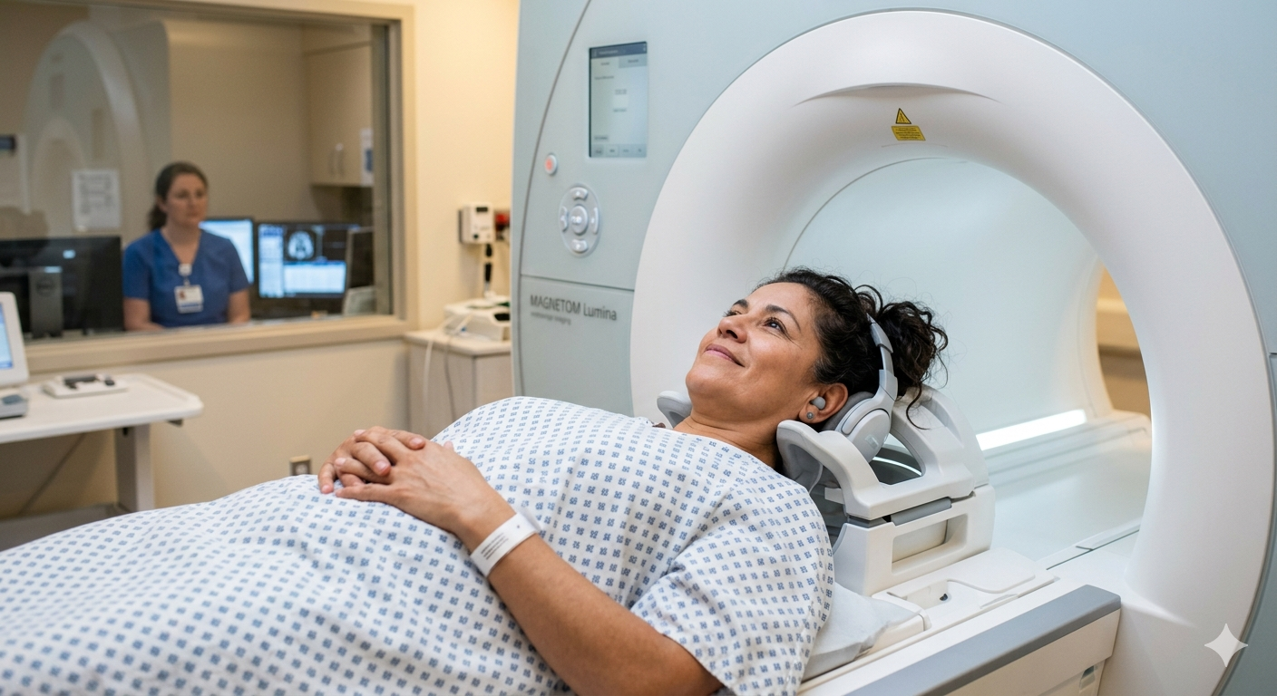
MRI (more detailed scan)
An MRI gives a more detailed picture of the pelvis.
It can help find deeper endometriosis, especially in areas like the bowel, bladder, or behind the uterus.
Often used when symptoms are severe or surgery is being planned.
Limitation: still may miss smaller areas.
Important to know
Imaging helps map and suspect endometriosis, but it cannot rule it out if results are normal.
The only way to definitively diagnose endometriosis is through surgery (laparoscopy), where a doctor looks inside and confirms it.
Simple way to think about it
Imaging is like a map—it can show the bigger or deeper areas of endometriosis, but it may not catch everything. That’s why symptoms and clinical expertise are just as important as the scan results.